
![]() Due to delay in AV node conduction
Due to delay in AV node conduction
![]() Differential Diagnosis of prolong PR interval:
Differential Diagnosis of prolong PR interval:
Increase vagal tone
Hyperkalemia
Digitalis
Ca and Beta Blockers
Ischaemic heart disease (especially Inferior MI)
Old age
![]() Case 1
Case 1
![]() Case 2
Case 2
![]() Case 3
Case 3
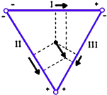
![]() Intermittent blocking of conduction through AV node or nerve pathways below.
Intermittent blocking of conduction through AV node or nerve pathways below.
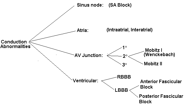
![]() Further Classified into 2 types, Mobitz I and II. ( I find this really confusing, so I rather think about where in the conduction system block occurs.)
Further Classified into 2 types, Mobitz I and II. ( I find this really confusing, so I rather think about where in the conduction system block occurs.)
![]() Mobitz type 1(Wenckebach)- Level of block is usually at AV node. There is progressive lengthening of the PR interval until the p wave is not conducted. The QRS complex is narrow; suggesting the level of block is the AV node.
Mobitz type 1(Wenckebach)- Level of block is usually at AV node. There is progressive lengthening of the PR interval until the p wave is not conducted. The QRS complex is narrow; suggesting the level of block is the AV node.
![]() Mobitz type 2- Level of block is below the AV node. Therefore PR interval is not prolong and remain constant. The width of QRS complex will usually indicates how far down the conduction is diseased. (ie. If block is very close to AV node QRS will be narrow, if QRS complex is wide then the block is further down the conduction system)
Mobitz type 2- Level of block is below the AV node. Therefore PR interval is not prolong and remain constant. The width of QRS complex will usually indicates how far down the conduction is diseased. (ie. If block is very close to AV node QRS will be narrow, if QRS complex is wide then the block is further down the conduction system)
![]() Case 1
Case 1
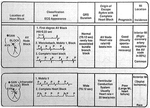
![]() Nothing gets through the AV node. Unrelated p waves and QRS complex (ie AV dissociation)
Nothing gets through the AV node. Unrelated p waves and QRS complex (ie AV dissociation)
![]() When block is at the level of the AV node, the takeover pacemaker is just below the node (His bundle), the QRS complex is therefore normal.
When block is at the level of the AV node, the takeover pacemaker is just below the node (His bundle), the QRS complex is therefore normal.
![]() When block develops in the infranodal conduction system, the takeover pacemaker is in the body of ventricule, the QRS is wide, and the rate is slow. This will be an indication for pacemaker therapy.
When block develops in the infranodal conduction system, the takeover pacemaker is in the body of ventricule, the QRS is wide, and the rate is slow. This will be an indication for pacemaker therapy.
![]() Case 1
Case 1
![]() Case 2
Case 2
![]() Case 3
Case 3
![]() When the right bundle branch is blocked, septum and left ventricle are activated normally. Current then spreads from left to the right ventricle, which is depolarised late.
When the right bundle branch is blocked, septum and left ventricle are activated normally. Current then spreads from left to the right ventricle, which is depolarised late.
![]() Features of RBBB:
Features of RBBB:
-Wide QRS duration of 120 ms or more
-RSR pattern in V1
-Slurring of S wave in left side leads (V4-6, I and aVL)
![]() Differential Diagnosis of RBBB:
Differential Diagnosis of RBBB:
Normal variant
ASD, Tetralogy of Fallot's
Acute, massive Pulmonary embolus
Cor pulmonale
![]() Case 1
Case 1
![]() Case 2
Case 2
![]() Case 3
Case 3
![]() Sequence of ventricular depolarisation is almost the opposite to RBBB.
Sequence of ventricular depolarisation is almost the opposite to RBBB.
![]() Features of LBBB:
Features of LBBB:
-Wide QRS complex in left side leads (I, aVL and V6), 120 ms or more
-Loss of septal q wave in left side leads (I, aVL and V6)
![]() Differential Diagnosis of LBBB:
Differential Diagnosis of LBBB:
Occaionally a normal variant
Ischaemic heart disease
Hypertension
Cardiomyopathy
Calcific aortic stenosis
Idiopathic fibrosis
Following cardiac surgery
![]() Case 1
Case 1
![]() Case 2
Case 2
![]() The left bundle divides into anterior and posterior branches.
The left bundle divides into anterior and posterior branches.
![]() Left anterior fascicular block is more common than posterior block because of its size and location.
Left anterior fascicular block is more common than posterior block because of its size and location.
![]() Features of LAFB:
Features of LAFB:
-Left axis deviation more than -30°
-r wave in all inferior leads (II, III and aVF)
-Absence of other causes of left axis deviation
![]() Features of LPFB:
Features of LPFB:
-Usually in significant left ventricular disease
-Right axis deviation between 90°-120°
-Initial q wave in leads II, III, aVF
-T wave inversion in inferior leads
-Absence of other causes of right axis deviation
![]() Case 1
Case 1
![]() RBBB plus block of one of the two branches of the left bundle. (ie. RBBB+LAFB or RBBB+LPFB)
RBBB plus block of one of the two branches of the left bundle. (ie. RBBB+LAFB or RBBB+LPFB)
![]() Diagnosis is simple: RBBB plus features of LAFB/LPFB
Diagnosis is simple: RBBB plus features of LAFB/LPFB
![]() Case 1
Case 1
![]() A combination of bifascicular block and first-degree AV Block.
A combination of bifascicular block and first-degree AV Block.
![]() Case 1
Case 1